 Sunita Somvanshi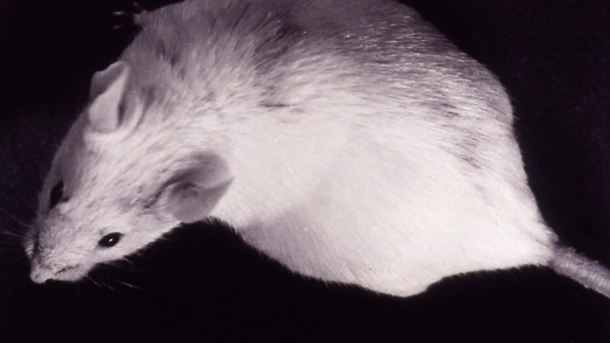
A team at Stanford Medicine has transplanted clusters of human brain cells into mice in a way that allowed the human tissue to become fully incorporated into the living animal’s neural architecture. The study, published in Nature on 16 September 2026, describes a method that solves one of the fundamental limits of laboratory brain research. The model may help researchers study human cortical development and neurological disease, but its usefulness for drug testing still requires further validation.
The technique is called xenocortication. Researchers transplanted human cortical organoids — small clusters of human brain cells grown from stem cells in a lab — into newborn mice whose cerebral cortex and hippocampus had been largely depleted beforehand. Creating that space in the developing mouse brain was the key methodological step. Previous organoid approaches faced limitations because tissue without a blood supply cannot grow beyond a small size. By transplanting during early mouse development, the Stanford team allowed the host animal’s own blood vessels to grow into the human tissue. The human organoids became vascularised — alive, connected, and functional.
The model may help researchers study disorders including autism, epilepsy and schizophrenia, but it is an experimental research tool at this stage, not a treatment platform. Earlier drug development has relied on brain models that lacked blood circulation and functional circuits.
At approximately three months after surgery, human-derived tissue accounted for approximately 91.9% of the cortical volume in the xenocortical mice. This does not mean that 91.9% of the entire brain was human. The human neurons grew, matured, established functional synapses with the host mouse’s neural circuits, and influenced sensory-motor behaviour.
Solving the Blood Supply Barrier in Brain Research
The vascularisation problem is the primary reason brain organoid research has, until now, remained limited in its clinical applications. A lab-grown brain cluster can develop the architecture of early human cortical tissue — layers of cells, early connections — but without a blood supply, cells at the centre cannot receive sufficient oxygen and nutrients. That is why organoid research has been constrained to studying early development rather than mature function.
The Stanford method sidesteps this by using the mouse’s own developmental biology to do the vascularisation work. The timing — transplanting into a newborn mouse whose brain is still in formation — means blood vessels grow into the human tissue as a matter of course. The result is tissue that can mature and remain alive for months.
The ethical framework for this research was reviewed under ISSCR guidance, Stanford’s Stem Cell Research Oversight committee and Institutional Animal Care and Use Committee processes. The study did not show evidence that the mice acquired human consciousness or human-like cognition; questions about neural chimeras remain an active area of ethical discussion.
The most immediate application is not a new treatment — it is a better research model. Research teams studying ALS, spinal muscular atrophy, schizophrenia, and epilepsy now have a model that integrates human cortical tissue into a living nervous system, which is where further research begins.
Did mice with human brain cells gain human consciousness?
The study did not show evidence that the mice developed human-like consciousness or cognition. The human neurons integrated into the mouse cortex and responded to sensory stimuli, but the transplanted tissue did not replicate human brain architecture, emotional capacity, or higher-order thinking. The research was reviewed under ISSCR guidance and institutional ethics oversight.