A breakthrough AI system called PanDerm is changing how doctors find and treat skin cancer. Developed by researchers from Monash University, University of Queensland, and Medical University of Vienna, this tool helps doctors spot dangerous skin changes earlier and more accurately.
PanDerm works by looking at different types of skin images at once – something no AI has done before. When doctors use PanDerm, their accuracy in diagnosing skin cancer improves by 11%. For healthcare workers who aren’t skin specialists, the improvement jumps to 16.5%.
“The tool could be particularly valuable in busy or resource-limited settings, or in primary care where access to dermatologists may be limited,” explains Professor H. Peter Soyer from the University of Queensland.
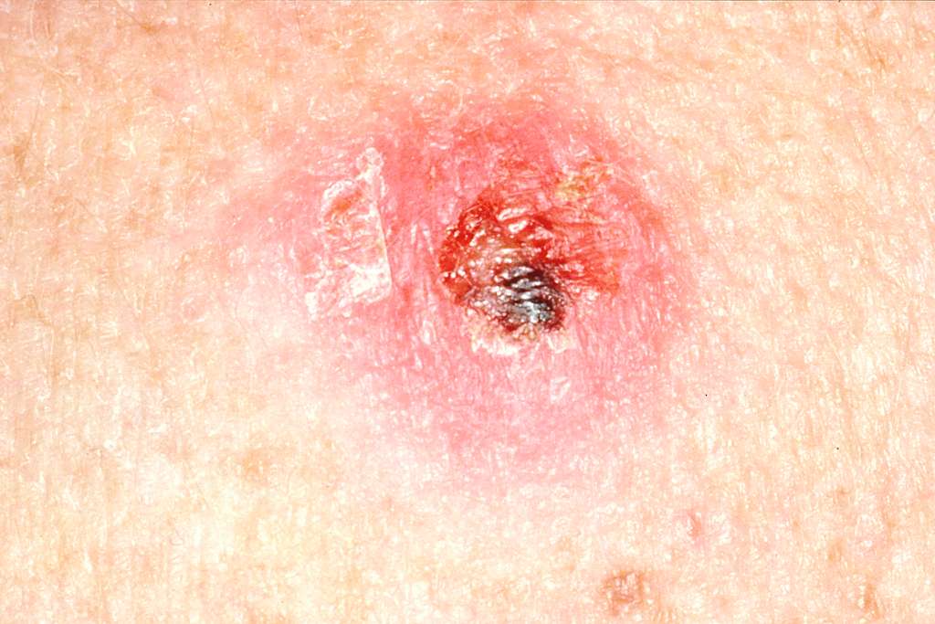
In clinics today, dermatologists fit PanDerm into their regular process. They take the usual pictures of concerning skin spots – close-ups, special magnified images, and sometimes full-body photos. PanDerm then analyzes these images together and gives doctors a second opinion about what they’re seeing. This helps spot subtle changes that might signal early skin cancer.
Similar Post
The need for better detection is urgent. About 70% of people worldwide have some type of skin condition. There are over 3,000 different skin diseases doctors must identify. In the U.S. alone, doctors diagnose about 9,500 people with skin cancer daily, and more than two people die from it every hour.
When skin cancer is found early, treatment works better. PanDerm helps by checking if a spot might be cancerous, tracking changes in moles over time, counting moles, and even predicting if a cancer might come back after treatment.
Professor Victoria Mar, Director of Alfred Health Victorian Melanoma Service, points out: “This kind of assistance could support earlier diagnosis and more consistent monitoring for patients at high risk of melanoma.”

The system learned from over two million skin images from 11 institutions across multiple countries. This diverse training helps it work well for different skin types and in various healthcare settings.
PanDerm doesn’t replace doctors – it helps them. By processing complex images quickly, it gives doctors more confidence in their decisions and helps them use their time more efficiently. This could mean faster appointments and shorter waiting times for patients.
The tool is still being tested before wider use in hospitals and clinics. Researchers continue to improve it to handle more skin conditions and work well for all patients.
The research appears in the journal Nature Medicine.
Frequently Asked Questions
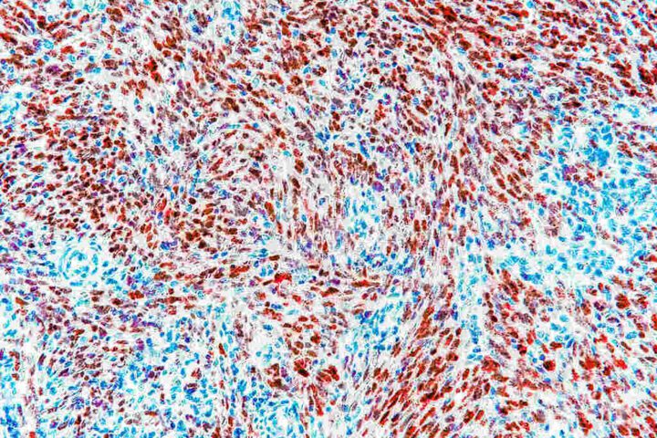
PanDerm is an AI-powered tool that analyzes multiple types of medical images simultaneously to detect skin cancer and other skin conditions. Unlike previous AI systems that were limited to single tasks, PanDerm can process close-up photos, dermoscopic images, pathology slides, and total body photographs together. This approach mimics how dermatologists work, synthesizing information from different visual sources to make more accurate diagnoses.
Research shows that PanDerm improved skin cancer diagnosis accuracy by 11% when used by dermatologists. For healthcare workers who aren’t skin specialists, the improvement was even greater, boosting diagnostic accuracy for various skin conditions by 16.5%. Remarkably, PanDerm can also detect concerning skin changes before they become visible to human clinicians, potentially enabling earlier intervention.
No, PanDerm is designed as a support tool, not a replacement for human clinicians. It helps doctors process and interpret complex imaging data more efficiently and accurately. This is particularly valuable in fast-paced or resource-limited settings where dermatologists may be scarce. PanDerm functions alongside clinicians, offering assistance in analyzing the spectrum of skin images that doctors routinely use.
Skin conditions affect approximately 70% of the global population, with over 3,000 different skin diseases identified by dermatologists. In the United States alone, about 9,500 people are diagnosed with skin cancer every day, and more than two people die from it every hour. Early detection is crucial for effective treatment, which is why tools like PanDerm that improve diagnostic accuracy are so important.
PanDerm was trained on an expansive dataset of over two million skin images meticulously curated from 11 different institutions across multiple countries. This diverse training dataset ensures a high degree of reliability across different patient demographics, imaging equipment, and lesion types. The international collaboration behind PanDerm helps ensure it works well across various healthcare systems worldwide.
Currently, PanDerm is in the evaluation phase before broader healthcare implementation. While the research results are promising, the team is working to develop more comprehensive evaluation frameworks that address a wider range of dermatological conditions and clinical scenarios. They’re establishing standardized protocols for cross-demographic assessments and investigating the model’s performance in varied real-world clinical settings.
